
I hope you have had a good start to 2024. I’ve had a great week, as my shoulder has been improving faster than I thought with physio, and I even managed 5 holes of golf yesterday with my son (playing with him is one of my all-time favourite things to do, and maybe I should injure myself more often – I was only 1 over par after 5).
In my video this week, I talk about the biological process of atherosclerosis, or ‘furring-up’ of heart arteries, and demonstrate this using a model.
This video should be up in the member’s area shortly, I hope you enjoy it.
However, as interesting as this is, what patients really want to know is whether or not they have any issues. The good news is that this is now easy with a special CT scan, so I thought I would say a bit more about them. One of my reasons for this was a patient I saw this week.
CT coronary angiogram scans take only a matter of seconds (in fact – just a single heart beat), have a low radiation dose (on a good scanner such as we are fortunate to have in Bournemouth) equivalent to a year’s background radiation, and are painless – no tubes going into the heart.
My patient was in her early 80s, and when I got the CT scan back, was a little surprised that there was no evidence of atherosclerosis whatsoever! I would estimate I see this in <5% of cases at this age. Good for her. The difficulty is, whilst usually more risk factors = more heart disease, this case just demonstrates it can be hard to tell. I could also cite many examples of younger patients where a CT scan unexpectedly demonstrated furring up of the heart arteries.
The image below shows the coronary arteries in the lighter colour, sitting on top of the reddish brown heart muscle. This is a 3D scan, but we actually use 2D greyscale images to make a diagnosis. If we do see evidence of atherosclerosis, the technology at this stage is such that we can tell whether it has any high risk or low risk features! Clever stuff.
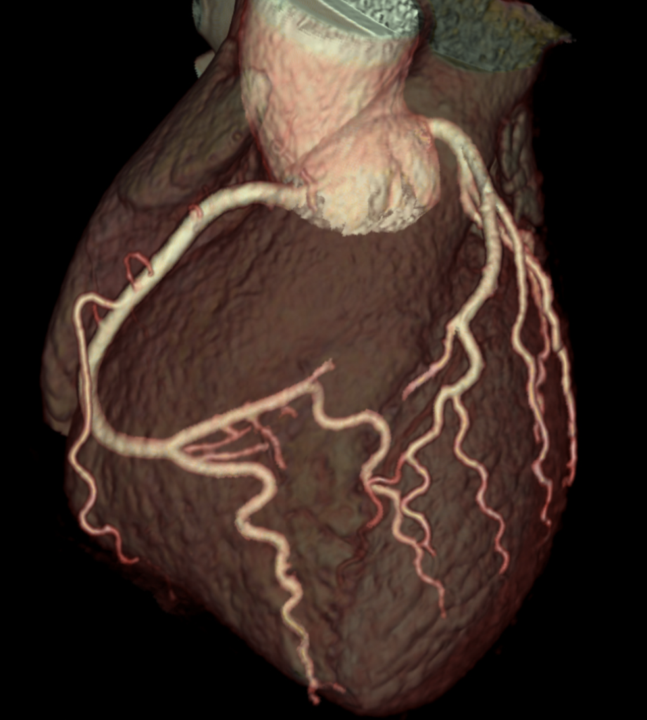
You will notice in the image above that the arteries are rather ‘wiggly’ (the medical term we use is tortuous). This is more common in patients with high blood pressure. We also get additional information such as the size and shape of the aorta (the body’s main blood vessel), the presence of any clots within the heart, and how the lungs and tissues around the heart look in addition. Lastly, CT scanning is a good way to image the pericardium, or ‘bag’ that the heart sits in.
I’m on call this week, and I would anticipate seeing a number of patients in whom a CT scan will play a key part of the diagnostic process. It’s great to be able to perform a scan which is so quick and low risk.
I hope you have a good week, I’m anticipating a busy one! I hope also if you made any new year’s resolutions that they are going well!